
Which Surgical Instruments Are Used to Close Off Blood Vessels
Controlling bleeding during surgery—vascular control—is a fundamental skill that impacts patient safety, operative efficiency, and overall outcomes. The process of closing off blood vessels involves a wide array of specialized tools and techniques, from simple mechanical instruments to advanced energy-based devices. This article provides a comprehensive overview of the instruments used for vascular closure, organized by technique category and enriched with practical details, tables, and relevant citations.
Importance of Vascular Control in Surgery
Effective vascular control is essential in all surgical fields to maintain a clear operative field and to safeguard patient well-being.
Excessive bleeding can lead to impaired visualization, prolonged operative times, and increased risk of postoperative complications such as hematoma or infection.
Achieving hemostasis stabilizes the patient’s hemodynamic status, reduces the need for blood transfusions, and shortens hospital stays.
Suboptimal vascular control may elevate morbidity and mortality, underscoring the need for reliable instruments and techniques.
Primary Techniques for Closing Blood Vessels

Surgeons rely on four main categories to stop blood flow:
- Mechanical Methods
- Chemical Methods
- Thermal/Energy-Based Methods
- Adjunct Techniques
Each category comprises various instruments and approaches, selected based on vessel size, location, and clinical context.
1. Mechanical Methods
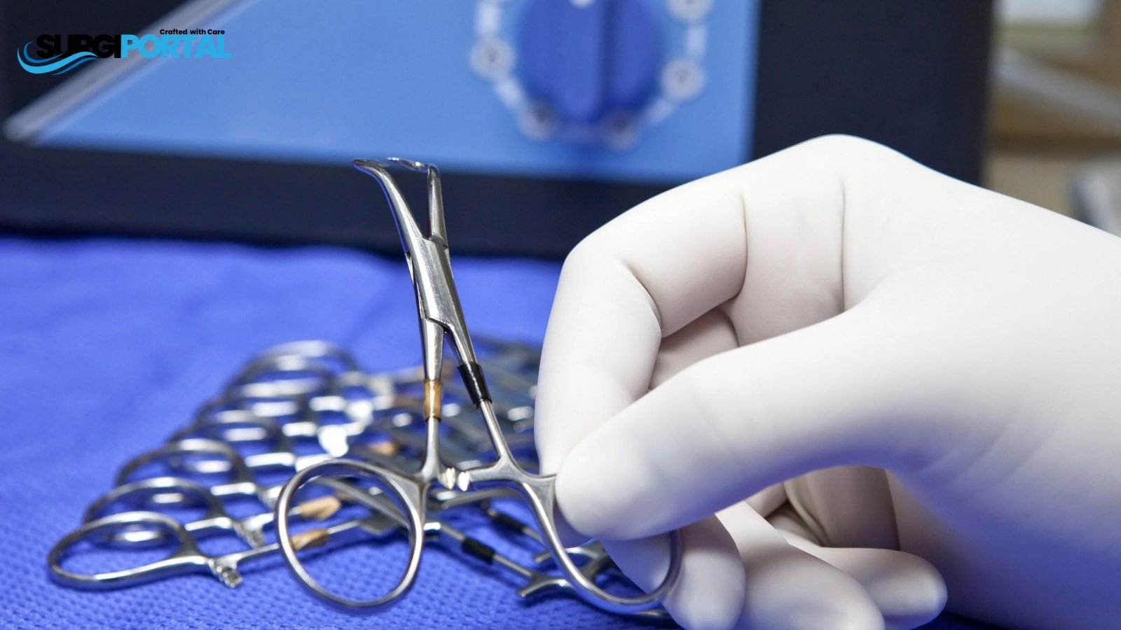
1.1 Hemostatic Forceps (Hemostats)
Hemostats—also known as artery forceps—are the cornerstone of mechanical hemostasis. They grasp and temporarily occlude vessels to permit suture ligation or cautery.

Key characteristics include serrated jaws, a locking ratchet, and availability in straight or curved designs:
- Kelly Forceps (Standard Kelly): Medium-sized, straight or curved; used for vessels up to 4 mm.
- Mosquito Forceps: Small, fine-tipped hemostats; ideal for capillaries or venules up to 2 mm.
- Crile Forceps: Fully serrated jaws for robust tissue or vessel control.
- Halsted Forceps: Similar to mosquito hemostats but slightly larger.
Table 1 summarizes common hemostats:
| Instrument | Jaw Pattern | Vessel Size (mm) | Use Case |
|---|---|---|---|
| Kelly Forceps | Half-serrated | 2–4 | General vessel occlusion |
| Mosquito Forceps | Fine serrations | ≤2 | Microvascular, capillary bleeding |
| Crile Forceps | Fully serrated | 3–6 | Ligating larger vessels or tissues |
| Halsted Forceps | Fine serrations | ≤2 | Delicate, microvascular procedures |
1.2 Vascular Clamps

For larger arteries or when more precise flow control is needed, vascular clamps are used. Common varieties include:
- Bulldog Clamps: Small, spring-loaded clamps for microvascular control (vessels up to 3 mm).
- Satinsky Clamps: Curved clamps to occlude large vessels such as the inferior vena cava.
- Kocher Clamps (Ochsner): Heavy ratcheted clamps with interlocking teeth for tough tissue and vessels (up to 10 mm).
- Rochester-Carmalt Clamps: Longitudinal serrations ideal for pedicle and vascular bundle occlusion.
Table 2: Vascular Clamps Comparison
| Clamp Type | Opening Style | Vessel Size (mm) | Specialty Applications |
|---|---|---|---|
| Bulldog Clamp | Spring-loaded | ≤3 | Microsurgery, AV shunts |
| Satinsky Clamp | Curved jaws | 5–15 | Cardiovascular, large vessels |
| Kocher Clamp | Interlocking teeth | 4–10 | General, vascular surgery |
| Rochester-Carmalt | Longitudinal serr. | 6–12 | Gynecologic, large pedicles |
2. Chemical Methods
2.1 Topical Hemostatic Agents

Chemical agents promote coagulation when mechanical measures are impractical:
- Passive Agents (Absorbable Collagen, Gelatin, Oxidized Cellulose) provide a scaffold for clot formation.
- Active Agents (Thrombin products, e.g., bovine or recombinant) directly catalyze fibrin clot formation.
Oxidized regenerated cellulose (Surgicel®) and gelatin sponges (Gelfoam®) absorb blood and activate platelets, whereas topical thrombin converts fibrinogen to fibrin at the site, achieving hemostasis within minutes.
Table 3: Hemostatic Agents
| Agent Type | Example | Mechanism | Absorption Time |
|---|---|---|---|
| Passive | Surgicel® | Scaffold‐driven clot formation | 7–14 days |
| Passive | Gelfoam® | Gelatin matrix for platelet action | 4–6 weeks |
| Active | Thrombin (Rec.) | Enzyme-mediated fibrin formation | 10–20 days |
| Sealant | Tisseel® | Fibrinogen + thrombin sealant | 2–8 weeks |
2.2 Vascular Clips and Ligature Clips

Ligature clips (e.g., titanium or polymer) offer permanent vessel closure. Applied via clip appliers, they pin vessels shut and remain in tissue, eliminating the need for suture knots for small to medium vessels. Advantages include:
- Fast application in minimally invasive surgery
- Consistent closure force
- Low tissue trauma
3. Thermal/Energy-Based Methods
3.1 Electrocautery
Electrosurgery utilizes electric current to coagulate vessels:
- Monopolar Cautery (Bovie®) cuts and coagulates but carries the risk of collateral thermal injury.
- Bipolar Cautery (Forcep-style devices) confines energy between tips for precise coagulation.
Monopolar is useful for broad tissue effects; bipolar is preferred for vascular sealing near critical structures.
3.2 Vessel Sealing Devices
Advanced bipolar vessel sealers fuse vessel walls through radiofrequency energy combined with pressure (Transcollation™), sealing vessels up to 7 mm with minimal thermal spread (e.g., Aquamantys™, CoolSeal Trinity™). Benefits include:
- Rapid sealing (≈1–2 seconds)
- Minimal collateral thermal damage (<1 mm)
- No foreign materials left in tissue
4. Staplers and Clip Appliers

4.1 Linear Cutting Staplers
Linear staplers place two parallel staple rows and divide between them, ideal for GI, thoracic, and vascular resections (e.g., EndoGIA®, SEC+, etc.). They ensure rapid closure of large vessels at resection margins.
4.2 Circular Staplers
Circular staplers (e.g., EEA®, Proximal Anastomosis) create permanent circular anastomoses, commonly in colorectal and vascular bypass surgeries. These instruments deliver consistent staple rings for secure luminal joining.
5. Sutures, Ligatures, and Vessel Loops
5.1 Sutures and Ligatures
Non-absorbable sutures (e.g., polypropylene, silk) provide long-term vascular support, whereas absorbable sutures (e.g., PDS, Vicryl®) degrade in weeks/months—ideal for temporary closures such as dural or GI repairs.
5.2 Vessel Loops
Vessel loops—silicon or latex bands—encircle vessels to achieve temporary occlusion during dissection or anastomosis. Their flexibility and color-coding aid rapid identification in complex fields.
6. Specialty-Specific Vascular Instruments
Different surgical fields employ tailored vascular tools:
- Neurosurgery: Micro-forceps, aneurysm clips
- Cardiac Surgery: Aortic cross-clamps, Debakey bulldogs
- Vascular Surgery: Satinsky clamps, Fogarty shunts (temporary perfusion)
- Orthopedics: Bone wax applicators, bone clamps for hemostasis
Each specialty integrates base techniques with unique instruments to optimize safety and precision.
7. Hemostatic Adjuncts: Topical Agents and Shunts
7.1 Temporary Vascular Shunts
Shunts (e.g., Argyle®, Javid®) maintain distal perfusion in trauma and reconstruction, allowing time for definitive repair. They prevent ischemia when vessel clamps cannot achieve prolonged flow control.
7.2 Topical Hemostatic Adjuncts
Adjuvants like tranexamic acid, epinephrine sprays, and gelatin-thrombin matrices complement mechanical and electrosurgical methods for diffuse oozing or surface bleeding.
8. Material and Design Considerations
Instrument material impacts performance and safety:
- Stainless Steel (420, 316L): Durable, corrosion-resistant
- Titanium: Lightweight, non-magnetic, ideal for MRI suites
- Polymeric Coatings: Reduce tissue adhesion, enhance sterility
Design aspects (ergonomic handles, smooth joint action, clear labeling) improve user comfort and reduce OR fatigue.
9. Instrument Sterilization and Maintenance

Proper processing ensures patient safety and instrument longevity:
- Cleaning: Manual or ultrasonic prior to sterilization
- Sterilization: Steam autoclave (121–134 °C)
- Inspection: Check for damage, corrosion, or blade dullness
- Maintenance: Routine lubrication of hinged instruments; periodic sharpening of scissors and knives.
10. Cost, Availability, and Procurement Factors
When selecting vascular instruments for a surgical inventory, consider:
- Initial Cost vs. Lifecycle Cost: Reusable vs. disposable impact on long-term budgets
- Procurement: Vendor reliability, supply chain resilience
- Compatibility: Integration with existing equipment and disposables
- Regulatory Compliance: ISO 13485, FDA 510(k), CE Mark approvals
Strategic sourcing and standardization can reduce OR tray redundancies by up to 60%, delivering substantial savings and efficiency improvements.
11. Emerging Trends and Innovations
- Robotic Vessel Sealing: LigaSure™ RAS on Hugo™ RAS system for advanced hemostasis in minimally invasive procedures8.
- Bioengineered Sealants: Autologous fibrin adhesives and synthetic hydrogels for customized tissue sealing.
- 3D-Printed Instrumentation: Patient-specific clamps and retractors for unique anatomies.
- Telesurgery: Real-time remote vascular interventions with precision instrument control.
These advancements aim to further minimize blood loss, reduce complications, and enhance surgical precision.
Conclusion
Closing off blood vessels is a multifaceted challenge requiring a blend of mechanical, chemical, and energy-based techniques. From hemostats and vascular clamps to staplers, clip appliers, vessel sealing devices, and biological adjuncts, each tool plays a pivotal role in achieving safe and efficient vascular control. Critical considerations include instrument material, ergonomics, cost, and sterilization methods. Emerging technologies such as robotic vessel sealing and advanced biosealants promise to redefine vascular closure in surgery. A thorough understanding of these instruments and strategies is essential for surgical teams striving to optimize outcomes while conserving resources and enhancing patient care.